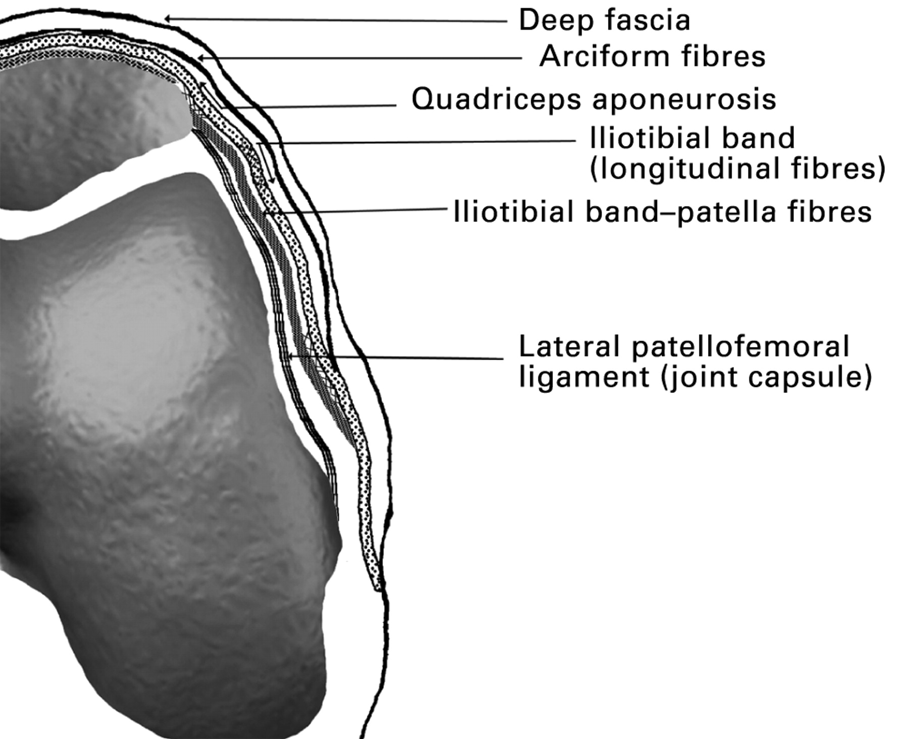
Anatomical Structure of Medial Retinaculum

The medial retinaculum is a fibrous band that forms the palmar (palmar) border of the carpal tunnel. It originates from the pisiform bone and triquetrum, and inserts onto the hamate bone and the base of the fifth metacarpal. The medial retinaculum is a key component of the carpal tunnel, which is a narrow passageway through which the median nerve and tendons of the flexor digitorum superficialis and profundus muscles pass.
Attachments
The medial retinaculum has two main attachments:
- Proximal attachment: The medial retinaculum originates from the pisiform bone and triquetrum, two bones located on the ulnar side of the wrist.
- Distal attachment: The medial retinaculum inserts onto the hamate bone and the base of the fifth metacarpal, two bones located on the radial side of the wrist.
Variations, Medial retinaculum
There are a number of variations in the anatomy of the medial retinaculum. These variations can be clinically significant, as they can affect the risk of developing carpal tunnel syndrome.
- The medial retinaculum may be thicker or thinner than normal. A thicker medial retinaculum can put pressure on the median nerve and tendons, increasing the risk of carpal tunnel syndrome.
- The medial retinaculum may be attached more proximally or distally than normal. This can also affect the risk of carpal tunnel syndrome, as it can change the angle at which the median nerve and tendons pass through the carpal tunnel.
- The medial retinaculum may have a hole or opening in it. This is known as a “transverse carpal ligament perforation,” and it can allow the median nerve and tendons to pass through the carpal tunnel without being compressed.
Pathophysiology of Medial Retinaculum Conditions

The medial retinaculum, a fibrous band that forms the roof of the carpal tunnel, can develop various conditions that affect its structure and function. These conditions, including thickening, inflammation, and fibrosis, can lead to carpal tunnel syndrome and other debilitating disorders.
Causes and Risk Factors
Several factors can contribute to the development of medial retinaculum conditions:
- Biomechanical Factors: Repetitive hand and wrist movements, such as typing, using hand tools, or playing musical instruments, can strain the medial retinaculum, leading to thickening and inflammation.
- Systemic Diseases: Conditions like diabetes, rheumatoid arthritis, and hypothyroidism can cause swelling and inflammation, which can affect the medial retinaculum.
- Age and Genetics: The medial retinaculum naturally thickens with age, and certain genetic factors may increase the risk of developing conditions.
Mechanisms Leading to Carpal Tunnel Syndrome
Carpal tunnel syndrome, a common condition caused by medial retinaculum dysfunction, occurs when the median nerve becomes compressed within the carpal tunnel. The thickened and inflamed retinaculum narrows the tunnel, putting pressure on the nerve and causing symptoms like pain, numbness, and tingling.
Clinical Evaluation and Management of Medial Retinaculum Conditions

The evaluation and management of medial retinaculum conditions involve a combination of physical examination, diagnostic tests, and treatment options. Here’s a comprehensive guide to help healthcare professionals assess and address these conditions effectively.
Physical Examination
- Inspect the wrist for swelling, tenderness, and any visible deformities.
- Palpate the medial retinaculum for tenderness or thickening.
- Perform the Tinel’s sign test by tapping over the median nerve at the wrist crease; a positive result indicates nerve compression.
- Assess range of motion, grip strength, and sensory function in the median nerve distribution.
Diagnostic Tests
In addition to physical examination, diagnostic tests can help confirm the diagnosis of medial retinaculum conditions:
- Nerve conduction studies (NCS) measure the electrical activity of the median nerve and can detect nerve compression.
- Electromyography (EMG) assesses the electrical activity of muscles innervated by the median nerve, helping to determine the severity of nerve damage.
- Magnetic resonance imaging (MRI) provides detailed images of the wrist and can reveal structural abnormalities, such as thickening or inflammation of the medial retinaculum.
Differential Diagnosis
Carpal tunnel syndrome is the most common medial retinaculum condition, but other conditions can mimic its symptoms. The differential diagnosis includes:
- Guyon’s canal syndrome (compression of the ulnar nerve in the wrist)
- Pronator teres syndrome (compression of the median nerve in the forearm)
- Flexor carpi radialis tendinitis (inflammation of the flexor carpi radialis tendon)
- Ganglion cysts (fluid-filled sacs that can compress the median nerve)
Treatment Options
The treatment of medial retinaculum conditions depends on the severity of the condition and the patient’s individual needs.
Non-Surgical Treatment
- Splinting to immobilize the wrist and reduce pressure on the median nerve.
- Corticosteroid injections to reduce inflammation and pain.
- Physical therapy to improve range of motion, strengthen muscles, and reduce nerve compression.
Surgical Treatment
If non-surgical treatment fails to provide relief, surgery may be necessary. The most common surgical procedure is carpal tunnel release, which involves cutting the medial retinaculum to relieve pressure on the median nerve.
Surgery is typically successful in relieving symptoms, but it may take several weeks or months for full recovery. Physical therapy is often recommended after surgery to restore range of motion and strength.
The medial retinaculum, a band of connective tissue in the wrist, serves as a vital support structure. Its intricate network of fibers weaves together like a celestial tapestry, guiding tendons and protecting delicate nerves. Much like the seamless transition between scenes in theresa randle bad boys 4 , the medial retinaculum orchestrates a symphony of movement, ensuring the wrist’s effortless grace.
The medial retinaculum, a ligament that stabilizes the wrist joint, is a fascinating structure. Its intricate composition and function are similar to the delicate balance of life and death, as exemplified by the untimely demise of basketball legend Jerry West.
How did Jerry West die ? His passing was a somber reminder of the fragility of life, just as the medial retinaculum serves as a delicate yet essential support for our wrists.
The medial retinaculum, a thick band of connective tissue in the wrist, plays a crucial role in stabilizing the carpal bones. Like Jerry Weat , a renowned orthopedic surgeon, the medial retinaculum ensures the proper alignment and movement of the wrist joint.
Its presence prevents excessive bending or twisting, safeguarding the delicate structures within the wrist from potential injury.
Like the medial retinaculum, which binds the tendons of the flexor muscles in the wrist, the connection between Jennifer Hudson and Common is unbreakable. Their shared experiences, from love to heartbreak, have woven an enduring bond that reflects the resilience and adaptability of the medial retinaculum itself.
The medial retinaculum, a fibrous band that holds tendons in place, serves as a reminder of the importance of healthy habits. Just as a recalled soft drink can be detrimental to our well-being, neglecting our physical health can lead to ailments.
The medial retinaculum, a vital component of our anatomy, deserves our attention, reminding us to make conscious choices for a fulfilling life.
